


Principles of photoacoustic imaging
Photoacoustic Imaging (PAI) is a medical imaging technique that combines optics and acoustics to generate ultrasonic signals using the interaction of light with tissue to obtain high-resolution tissue images. It is widely used in biomedical fields, especially in tumor detection, vascular imaging, skin imaging and other fields.
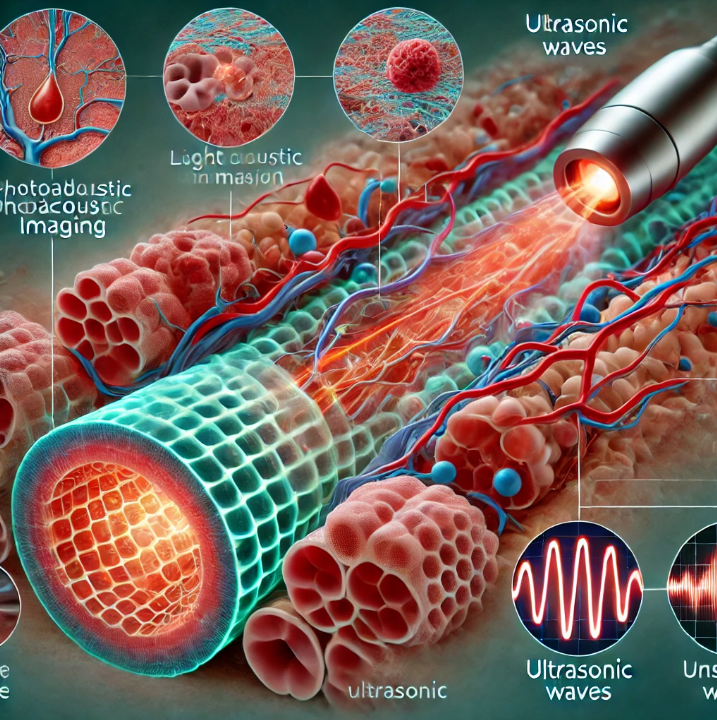
Principle:
1. Light absorption and thermal expansion: – Photoacoustic imaging uses the thermal effect produced by light absorption. The pigment molecules in the tissue (e.g., hemoglobin, melanin) absorb photons (usually near-infrared light), which are converted into heat energy, causing local temperatures to rise.
2. Thermal expansion causes ultrasound: – Temperature rise leads to tiny thermal expansion of the tissue, which produces pressure waves (i.e. ultrasound).
3. Ultrasonic detection: – The generated ultrasonic waves propagate within the tissue, and these signals are subsequently received and recorded by ultrasonic sensors (such as ultrasonic probes).
4. Image reconstruction: the collected ultrasonic signal is calculated and processed to rebuild the structure and function image of the tissue, which can provide the optical absorption characteristics of the tissue. Advantages of photoacoustic imaging: High contrast: Photoacoustic imaging relies on the light absorption characteristics of tissues, and different tissues (such as blood, fat, muscle, etc.) have different abilities to absorb light, so it can provide high-contrast images. High resolution: Using the high spatial resolution of ultrasound, photoacoustic imaging can achieve millimeter or even sub-millimeter imaging accuracy. Non-invasive: Photoacoustic imaging is non-invasive, light and sound will not cause tissue damage, very suitable for human medical diagnosis. Depth imaging capability: Compared with traditional optical imaging, photoacoustic imaging can penetrate several centimeters under the skin, which is suitable for deep tissue imaging.
Application:
1. Vascular imaging: – Photoacoustic imaging can detect the light-absorbing properties of hemoglobin in the blood, so it can accurately display the structure and oxygenation status of blood vessels for monitoring microcirculation and judging diseases.
2. Tumor detection: – Angiogenesis in tumor tissues is usually extremely abundant, and photoacoustic imaging can help early detection of tumors by detecting abnormalities in vascular structure.
3. Functional imaging: – Photoacoustic imaging can assess the oxygen supply of tissues by detecting the concentration of oxygenation and deoxyhemoglobin in tissues, which is of great significance for the functional monitoring of diseases such as cancer and cardiovascular disease.
4. Skin imaging: – Because photoacoustic imaging is very sensitive to superficial tissue, it is suitable for early detection of skin cancer and analysis of skin abnormalities.
5. Brain imaging: Photoacoustic imaging can obtain cerebral blood flow information in a non-invasive manner for the study of brain diseases such as stroke and epilepsy.
Challenges and development directions of photoacoustic imaging:
Light source selection: Light penetration of different wavelengths is different, how to choose the right wavelength balance resolution and penetration depth is a challenge. Signal processing: The acquisition and processing of ultrasonic signals require high-speed and accurate algorithms, and the development of image reconstruction technology is also crucial. Multimodal imaging: Photoacoustic imaging can be combined with other imaging modalities (such as MRI, CT, ultrasound imaging) to provide more comprehensive biomedical information.
Photoacoustic imaging is a new and multi-functional biomedical imaging technology, which has the characteristics of high contrast, high resolution and non-invasive. With the development of technology, photoacoustic imaging has broad application prospects in medical diagnosis, basic biology research, drug development and other fields.
Post time: Sep-23-2024